USTC Realizes Single-pixel Imaging of Single Living Cells
A research team led by Prof. GONG Lei from the University of Science and Technology (USTC) and collaborators developed a three-dimensional single-pixel imaging (3D-SPI) approach based on 3D light-field illumination(3D-LFI), which enables volumetric imaging of microscopic objects with a near-diffraction-limit 3D optical resolution. They further demonstrated its capability of 3D visualization of label-free optical absorption contrast by imaging single algal cells in vivo. The study titled “Optical Single-Pixel Volumetric Imaging by Three-dimensional Light-Field Illumination” was published in Proceedings of the National Academy of Sciences (PNAS) on July 25.
SPI has become an attractive 3D imaging modality. Through single-pixel detectors instead of conventional array sensors, the performance of SPI exceeds the conventional ones in spectral range, detection efficiency, and timing response. Furthermore, the single-cell cameras outperform conventional imaging methods at weak intensity, single-photon level, and precise timing resolution.3D-SPI techniques generally depend on time-of-flight (TOF) or stereovision to extract depth information. However, existing implementations can only reach a millimeter level at best, which is incapable of imaging microscopic objects like cells.
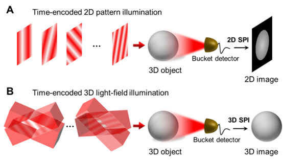
Schematic diagram of 3D-SPI technique (Image by LIU Yifan)
To exceed the resolution limitation, the researchers built a 3D-LFI-SPM prototype. As a result, the prototype achieves an imaging volume of ~390×390×3,800 μm3 and a resolution of up to 2.7 μm laterally and 37 μm axially. They performed label-free 3D imaging of living Haematococcus pluvialis cells and successfully counted the living cells in situ.
Predictably, the approach can be applied to visualize various absorption contrasts of biological samples. With depth-resolved imaging ability, scientists might be potentially able to monitor cell morphology and growth in situ in the future. The research opens the door to high-performance 3D SPI with applications in biomedical research and optical sensing.
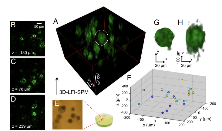
Label-free volumetric imaging of living H. pluvialis cells. (Image by LIU Yifan)
(Written by WENG Guchen edited by SHU Yukang, USTC News Center)
Back